Chipping Away at Brain Cancer

Combining microchip engineering techniques with cutting-edge gene profiling, scientists at Columbia University have developed a new way to study drug responses in living slices of human brain tumor cells. The system, using a type of chip called a microfluidic device, has already revealed new details about how these aggressive tumors resist chemotherapy drugs and could help researchers develop more effective treatments.
The work grew from earlier efforts to study glioblastoma tumors removed from patients during surgery. “These samples that we’re getting from our colleagues who resect these tumors clinically, they’re alive, and we can actually do experiments directly on those surgical samples,” says Peter Sims, PhD, associate professor of systems biology at Columbia and senior author on the new study, which appears in the journal Lab Chip.
From petri dishes to microfluidic chip
Sims’s lab had previously developed culture techniques that allowed them to grow these tumor slices in petri dishes, where they could test the effects of various drugs on the cancer cells’ survival. “These tissue slices mimic all the different cell types that coexist in these tumors, so we used single cell sequencing to assess how the drug affected each cell type independently,” says Sims, who is also co-leader of the Precision Oncology and Systems Biology (POSB) program at the HICCC. As interesting as those findings were, he wanted to improve the system in order to automate the cells’ maintenance and get more quantitative results.
It took a research symposium to discover that help was just next door. “Sam Sia’s lab is literally across the hall from my lab, but we didn’t really have a deep scientific conversation until we were at a meeting in a different venue,” says Sims. Sia, a professor of biomedical engineering at Columbia, is one of the leading researchers in microfluidic device development. The two scientists quickly realized that putting explanted tumor slices into tiny chambers on a chip would give them precise, automated control over the cells’ microenvironments and yield better and more comprehensive data.
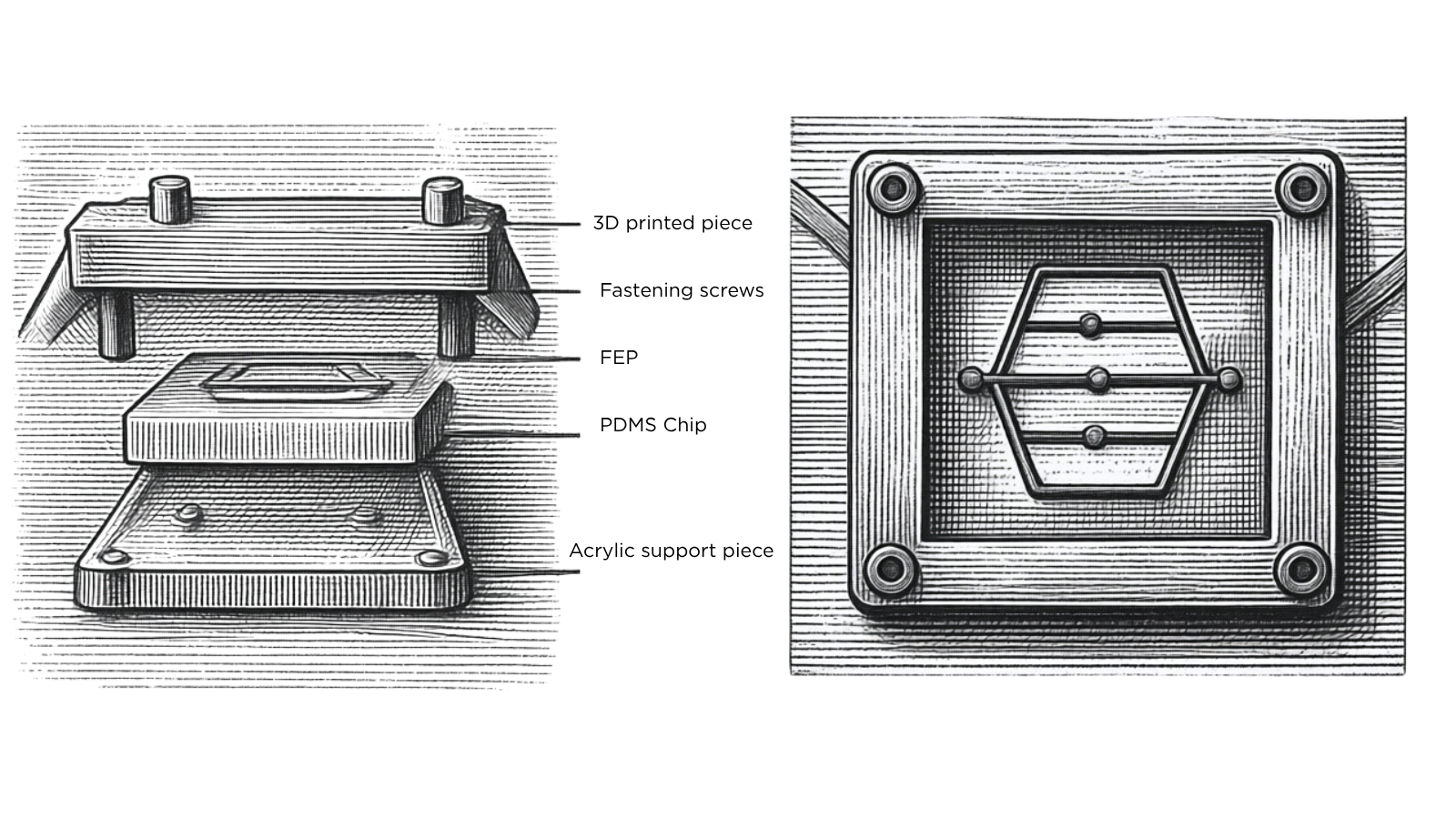
Digital rendering of a microfluidic device engineered to test drug responses in living brain tumor slices. The device accommodates five thick slices from a single tumor, each placed in an individual growth chamber for controlled microfluidic perfusion.
"In most organ-on-a-chip systems, the tissues are confined to small, enclosed regions and cannot be easily recovered,” says Sia. "Here, we developed a chip where the tissues could be perfused controllably with media or drugs, then taken out of the chip for powerful single-cell sequencing analysis."
Adding genomic insight to drug response
After several iterations to overcome technical challenges, the two labs developed devices that could hold five thick slices from a single tumor in individual growth chambers, while controlling the flows of nutrients and drugs to each. The system comes much closer to the conditions in which the tumors grow in a patient’s brain, compared to the team’s previous cultures.
The cells are under less oxidative stress, they’re more robust, they’re harder to kill, and I suspect we’re getting more realistic results from the experiments that are done in this way,
Importantly, unlike many microfluidic systems, the new chips are also designed so that the cells can be recovered from them alive later. “The cool feature of this device is that at the end of the experiment, you can retrieve the sample and then do a very sophisticated genomic analysis on it afterward,” says Sims. Using single-cell gene expression profiling is especially important for glioblastomas, which tend to contain multiple cell populations with different phenotypes. Even if a drug kills a majority of cells in a tumor, the surviving cells can start new tumors. The new study shows this pattern clearly, with sub-populations of cells exhibiting major differences in drug susceptibility.
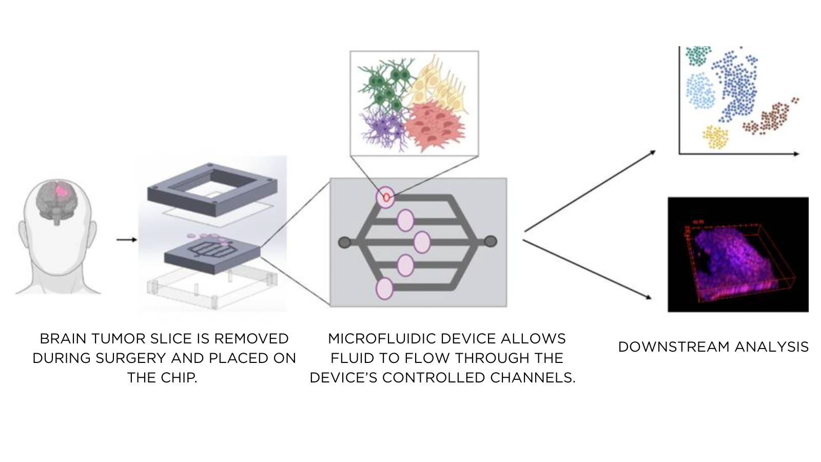
The new system for testing brain tumor drug responses, described in Lab on a Chip, places explanted tumor slices into small chambers on a microfluidic chip, enabling precise, automated control of the cellular microenvironment and generating more comprehensive data.
From the lab toward clinical trials
The two labs are already collaborating with others at Columbia to put the chips to work finding new therapies. “We’re actively using it on a daily basis in the lab to generate complementary preclinical data to the animal studies that are also ongoing at Columbia,” says Sims, adding that “we’d like to take that as a package to the FDA as stronger evidence to start a clinical trial in the future with the drugs that we are studying.”
