

Measuring Cancer Structure: New Topology-Based Biomarkers May Improve Breast Cancer Prediction

For decades, pathologists have diagnosed and graded breast cancer by looking at tissue samples under a microscope, searching for telltale signs of disorder in the structure of cells and tissues. Now, researchers at Columbia and their collaborators have developed a new computational approach that transforms those visual patterns into quantitative measurements, potentially improving how clinicians predict breast cancer outcomes and choose therapies.
In a recent study published in Cancer Research, researchers used mathematical tools known as topology to develop biomarkers quantifying the organizational structure of breast cancer tissue. The approach generated continuous numerical scores that predicted patient survival and treatment response more accurately than many traditional biomarkers, while also showing less variation across racial and ethnic groups.
Turning tumor structure into data
Cancer is often characterized by a loss of normal tissue organization. Healthy tissue forms structured, gland-like architectures, while malignant tissue becomes increasingly chaotic and disordered. Traditionally, pathologists assess these changes qualitatively through tumor grading systems.
“Pathologists have been looking at disorganized structure for years,” says Kevin Gardner, MD, PhD, chair of the department of pathology and cell biology at Columbia University Irving Medical Center and senior author of the study. “What topology allows us to do is move from something subjective to something quantitative.”
Topology is a branch of mathematics focused on shapes and spatial relationships. In this study, researchers applied a method called persistent homology to digital images of breast cancer tissue. Rather than simply identifying which cells were present, the method examined how tumor cells and immune cells were spatially organized relative to one another.
Using multiplex immunofluorescence imaging, the team mapped the precise coordinates of tumor cells, immune cells, and PD-L1 expression within tumor samples from more than 550 breast cancer patients from a racially diverse cohort in North Carolina. The researchers then used computational models to measure patterns of organization across the tissue.
The result was a set of topology-based biomarkers that could quantify tissue architecture on a continuous scale.
Predicting survival more accurately

The study found that these topology-based measurements strongly predicted breast cancer survival. Higher topology scores — indicating more organized tissue structure — were associated with longer survival and more favorable outcomes.
Importantly, the biomarkers outperformed several conventional approaches. Traditional biomarkers, including some gene and protein markers, often show variability in how accurately they predict outcomes across different patient populations. In contrast, the topology-based biomarkers remained highly predictive across both non-Hispanic Black and non-Hispanic white patient groups.
The researchers also integrated the topology measurements with gene expression data to create what they call topology-derived gene signatures. These signatures successfully predicted response to therapy in independent breast cancer clinical trial datasets.
“This work suggests that the spatial organization of the tumor contains important biological information that conventional biomarkers may miss,” says Jasmine McDonald, PhD, associate professor of epidemiology at Columbia and an author of the study.
Linking tumor structure and biology
Beyond prediction, the study also uncovered biological pathways associated with tumor disorganization. The researchers identified links between low topology scores and pathways involved in metabolism, immune suppression, and epithelial-to-mesenchymal transition — a process associated with cancer invasion and metastasis.
One particularly intriguing finding involved IL4-I1, a metabolic enzyme connected to aryl hydrocarbon receptor signaling, which has previously been implicated in tumor progression and immune regulation. The findings suggest that structural changes within tumors may be closely tied to metabolic and immune processes within the tumor microenvironment.
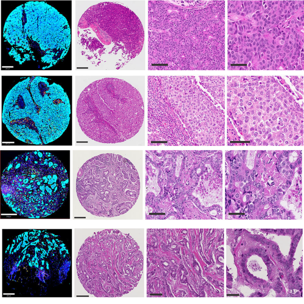
Multiplex immunofluorescent (left) and H&E-stained (right) breast tumor samples illustrating high versus low Base Topology Scores. Higher topology scores reflect more organized tissue architecture, while lower scores indicate greater structural disorganization. By quantitatively measuring these structural differences, the researchers developed biomarkers that predicted patient outcomes more accurately than many traditional approaches
A new frontier for digital pathology
The work reflects the growing convergence of digital pathology, computational biology, artificial intelligence, and mathematics in cancer research. By converting tissue architecture into quantitative data, researchers believe these methods could eventually complement existing pathology workflows and help guide treatment decisions.
The current study relied on multiplex immunofluorescence imaging, but the researchers are already working toward applying similar methods to standard pathology slides routinely used in clinics worldwide. Because those stains are inexpensive and already widely used, the approach could eventually help expand access to advanced cancer diagnostics far beyond major academic medical centers.
In the future, researchers envision a world in which tissue samples could be scanned digitally and analyzed using computational algorithms trained to recognize structural patterns linked to prognosis and therapeutic response — potentially making sophisticated pathology analysis more accessible in low-resource or geographically remote settings.
The long-term potential is that advanced cancer diagnostics could become faster, more quantitative, more accurate, and far more broadly accessible than they are today
While additional validation and clinical studies will be needed before the approach reaches routine clinical use, the findings highlight the potential for topology-based biomarkers to improve prognostic accuracy, deepen understanding of tumor biology, and potentially even predict therapeutic response in ways that are more consistent across diverse patient populations.
“Once you can put a number on an image, there’s a lot you can do,” Gardner says. “You can integrate it with genomics, clinical information, and other data streams to make more predictive models.”
While additional validation and clinical studies will be needed before the approach reaches routine clinical use, the findings highlight the potential for topology-based biomarkers to improve prognostic accuracy, deepen understanding of tumor biology, and potentially even predict therapeutic response in ways that are more consistent across diverse patient populations.
“For more than a century, pathology has relied on recognizing patterns visually,” Gardner says. “What’s exciting now is that we can begin to mathematically define those patterns and use them predictively. The long-term potential is that advanced cancer diagnostics could become faster, more quantitative, more accurate, and far more broadly accessible than they are today.”
References
The study includes investigators from the Herbert Irving Comprehensive Cancer Center across pathology, epidemiology, and systems biology, including Kevin Gardner, MD, PhD (member, Cancer Genomics and Epigenomics program); Hanina Hibshoosh, MD (member, Precision Oncology and Systems Biology program); Jasmine A. McDonald, PhD (associate director, Cancer Research Training and Education Core; member, Cancer Population Science program); and Raul Rabadan, PhD (co-leader, Cancer Genomics and Epigenomics program. Other Columbia University Irving Medical Center authors include Michael Miller, MD, PhD; Joy R. Winfield; Sai Tun Hein Aung, MD; Gustavo Martinez-Delgado, PhD; Ziv Frankenstein, PhD; Young-Ho Lee, PhD.
Related News
NIH Grant Supports Research to Improve Prediction of Advanced Breast Cancer Risk
Decoding Cancer: How Biomarkers Are Personalizing Treatment
Turning ‘Unknown’ Cancer Mutations into Personalized Treatment Strategies